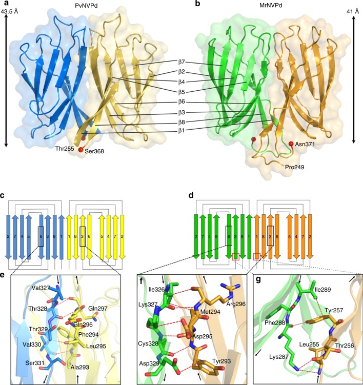
Fig. 3.
Topology and structural organization of the PvNVPd and MrNVPd. a, b Ribbon diagrams and surface presentations of the PvNVPd and MrNVPd are shown in similar orientations. The two C-termini are shown as red spheres. Each monomer of PvNVPd and MrNVPd is indicated in blue, yellow, green, and orange, respectively. c, d Schematic diagrams of the secondary structures in the PvNVPd and MrNVPd, respectively. Each monomer of PvNVPd and MrNVPd is indicated with colors as in a and b. e–g The hydrogen bonds, which stabilize the dimer, from specific β-strands β1, β3 and β6 with the key residues (sticks) at the interface are shown in red

