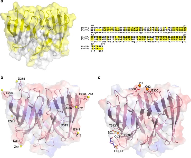
Fig. 4.
Insights on the surface of one dimeric MrNVPd. a Sequence-alignment variables between MrNVPd and PvNVPd mapped onto the surface of the P-domain. The hypervariable regions (yellow) from MrNVPd and PvNVPd are presented on the molecular surface and strictly variants residues are colored in yellow as well. b The molecular electrostatic potential surface of the dimeric MrNVPd with the Zn2+ ions. A side view of the dimeric MrNVPd is colored in red and blue for negatively and positively charged regions, and the Zn2+ ions are shown in yellow spheres. c The side view of the MrNVPd with the Cd2+ ions (orange spheres). The ligands of SO4 and HEPES are colored in yellow and blue, respectively

