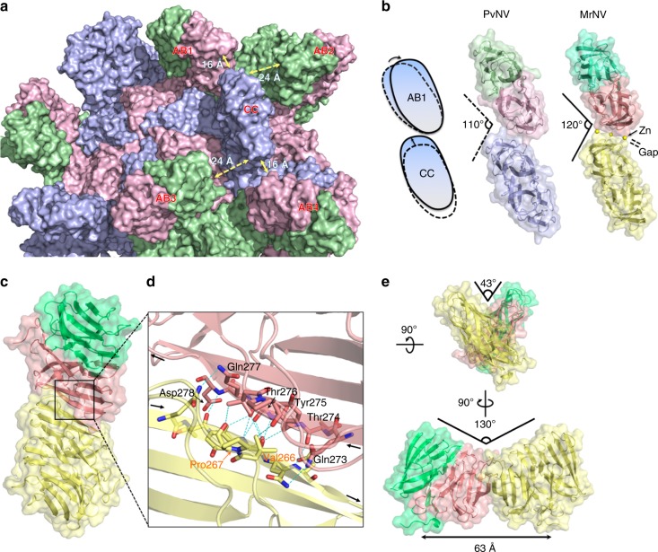
Fig. 5.
Intermolecular conformations of the dimeric P-domains. a The outer surface presentation of dimeric P-domains. The subunits A–C are colored in light green, pink, and purple, respectively. b The top views of the dimeric P-domains corresponding to the subunit-A/B and C/C dimers of the T = 3 PvNV-LP. The cartoon presentation of dimeric P-domains movement between A/B dimer and C/C dimer. The two PvNVPd dimers are engaged in the parallel conformation and are colored as subunits A–C in green, pink, and purple, respectively; the two MrNVPd dimers are engaged in the parallel conformation and the subunits A–C are colored in light green, salmon, and light yellow, respectively. The Zn2+ ions are located at the interface, resulting in a gap between two MrNVPd dimers. c A top view of the X-shaped conformation of the dimer–dimer MrNVPd. The two MrNVPd dimers are shown as blue and salmon, respectively. d The 2-fold dimerization interface of two MrNVPd dimers. The blue dotted lines indicate intermolecular hydrogen bonds between two MrNVPd dimers as colored in c. e The crossing angles of the two MrNVPd dimers. These two angles are stabilized by intermolecular hydrogen bonds, and are presented from two side views

