Figure 7.
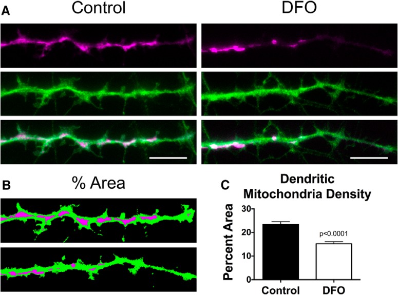
DFO decreases mitochondrial density in dendrite tips of 18 DIV neurons. Hippocampal neurons cultured from E16 mice were treated with DFO and 5-FU beginning at 3 DIV. At 18 DIV, mCherry-Mito-7 and GFP-expressing neurons were imaged with wide-field fluorescent microscopy and a 100× objective. A, Representative images of fluorescently labeled mitochondria (magenta) within terminal dendrites (green) and the corresponding merged images are shown for Control (left) and DFO (right) neurons. B, C, Dendrite and mitochondria images were thresholded and total area of each determined. The percentage of dendritic area taken up by mitochondrial area was calculated and expressed as the average percent area per neuron. The data from two independent cultures were pooled and are presented as mean ± SEM. Control: n = 36; DFO: n = 33 neurons. Student's t test was performed with α = 0.05; t(67) = 5.50.

