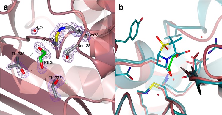
Figure 7.
Structure of the active site of BlaC S70C. (a) The crystal structure of BlaC S70C showing the presence of continuous 2mFo-DFc electron density connecting the side chains of Cys70 and Lys73. The proximity of the S and N atoms suggests the presence of a covalent sulfenamide bond. Positive difference mFo-DFc electron density was found in the active site of both protein chains in the crystal, which was modeled as polyethylene glycol (PEG). (b) Superposition of the X-ray crystal structures of BlaC S70C (pale crimson) and SHV-1 S70C mutant in complex with sulbactam (PDB 4FH2, teal). Sulbactam lies in the same position as the PEG molecule modeled in the active site of BlaC S70C.

