Fig. 3.
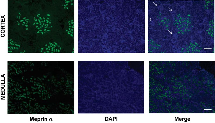
Renal expression of meprin-α. Images show meprin expression (green), nuclear staining with 4′,6′-diamidino-2-phenylindole (DAPI; blue), and merged images of the 2 in the cortical areas (top) and the medullary region (bottom). Arrows indicate structures that are compatible with the glomerulus in the cortex (scale bar, 200 µm).

