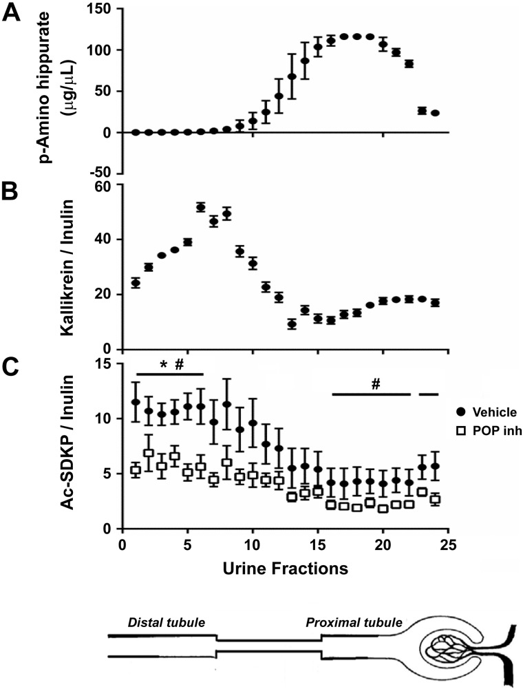
Fig. 6.
Quantitative analysis of N-acetyl-seryl-aspartyl-lysyl-proline (Ac-SDKP) in different nephron fractions through the stop-flow technique. A: PAH concentrations in consecutive urine fractions were analyzed to identify the fractions representing the proximal part of the nephron. B: kallikrein activity in consecutive urine fractions was analyzed to identify the fractions representing the distal part of the nephron. C: Ac-SDKP adjusted by water reabsorption along the nephron. Greatest amounts of Ac-SDKP are observed in the distal portion of the nephron, and smallest amounts are found in the proximal part of the nephron. Bottom curve (open squares) represents the Ac-SDKP-to-inulin ratio under infusion of the prolyl oligopeptidase enzyme inhibitor (POP inh) KYP-2047. Last 2 fractions in all panels represent the amounts of free flow (n = 7). *P < 0.01, distal nephron vs. proximal nephron. #P < 0.05, vehicle vs. POP inhibitor.

