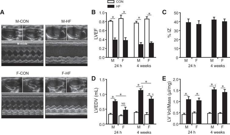
Fig. 1.
A: representative two-dimensional [2-D; top (diastole on the left and systole on the right)] and M-mode (bottom) echocardiographic images from control (CON) and heart failure (HF) rats from each sex at 4 wk after coronary artery ligation. B−E: quantitative comparison of echocardiographic parameters including left ventricular (LV) ejection fraction (LVEF; B), ischemic zone as a percentage of LV circumference (%IZ; C), LV end-diastolic volume (LVEDV; D), and LV volume-to-mass ratio (LV vol/mass; E) from CON and HF rats 24 h and 4 wk after coronary artery ligation. Data are means ± SE; n = 6–13 for each group. F, female; M, male; NS, not significant. *P < 0.05; §P < 0.05 vs. same group at 24 h.

