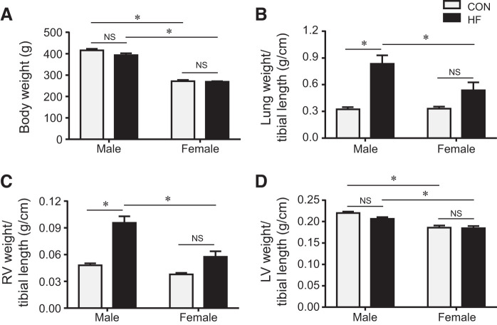
Fig. 2.
Quantitative comparison of anatomic parameters including body weight (A) and the ratios of lung weight (B), right ventricular (RV) weight (C), and left ventricular (LV) weight (D) to tibial length in control (CON) and heart failure (HF) rats of both sexes at 4 wk after coronary artery ligation. Data are means ± SE; n = 8–13 for each group. NS, not significant. *P < 0.05.

