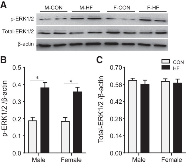
Fig. 7.

Representative Western blots (A) and quantitative comparison of protein expression for phosphorylated (p-) and total ERK1/2 (B and C) in the hypothalamic paraventricular nucleus in control (CON) and heart failure (HF) rats of both sexes at 4 wk after coronary artery ligation. Data are means ± SE; n = 5–6 for each group. F, female; M, male. *P < 0.05.
