Fig. 2.
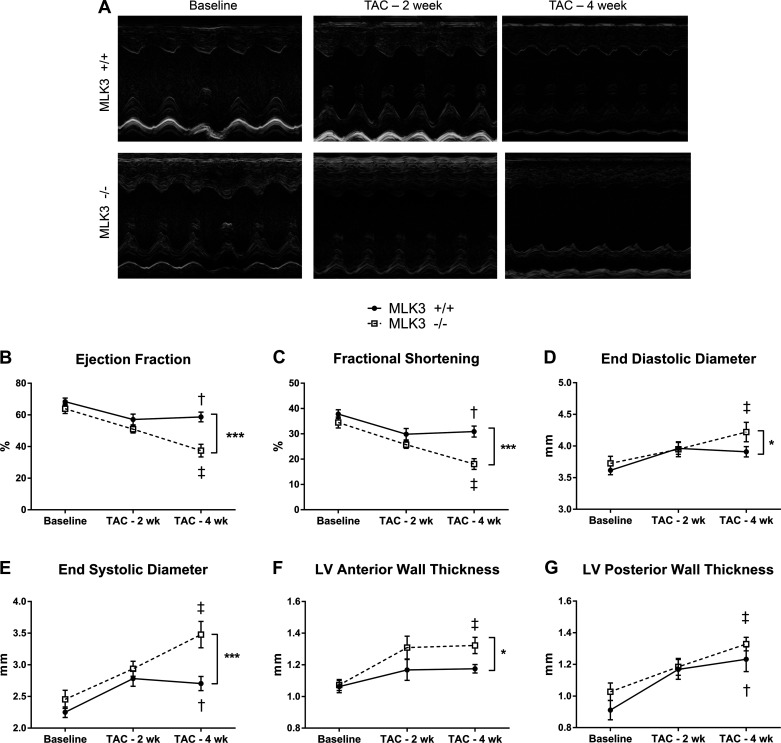
Mixed lineage kinase-3-deficient (MLK3−/−) mice develop increased left ventricular (LV) dilation and systolic dysfunction after 4 wk of pressure overload. Cardiac structure and function were evaluated by serial echocardiography in wild-type (MLK3+/+) and MLK3−/− mice subjected to sham or 25-gauge transverse aortic constriction (TAC) surgery for 4 wk. A: representative M-Mode images of the LV in MLK3+/+ and MLK3−/− mice at baseline and after 2 or 4 wk of TAC. B: ejection fraction calculated from M-Mode images of the LV. C: fractional shortening. D: end-diastolic diameter of the LV. E: end-systolic diameter of the LV. F: anterior LV wall thickness. G: posterior LV wall thickness; n = 8 per genotype. Data were analyzed by two-way ANOVA with repeated measures, and genotypes were compared using a Bonferroni posttest. †P < 0.05 baseline vs. 4-wk TAC for MLK3+/+ mice; ‡P < 0.05 baseline vs. 4-wk TAC for MLK3−/− mice; *P < 0.05 between genotypes at 4-wk TAC time point; ***P < 0.001 between genotypes at 4-wk TAC time point.

