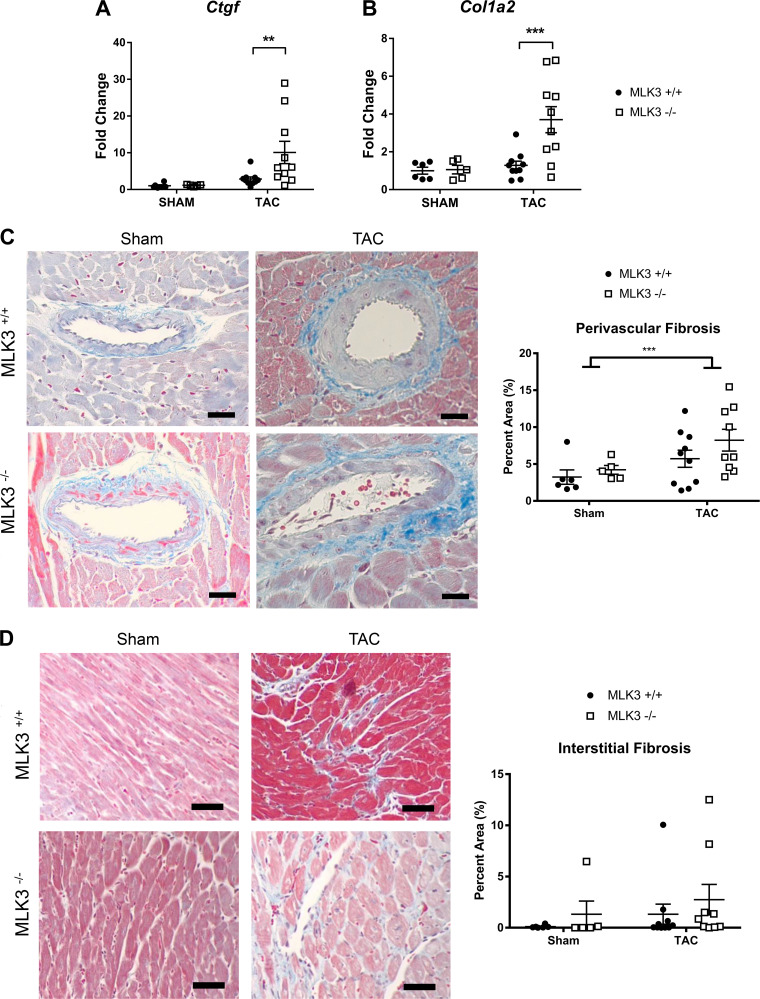
Fig. 4.
Analysis of profibrotic gene expression and cardiac fibrosis in mixed lineage kinase-3-deficient (MLK3−/−) left ventricles (LVs) after 4 wk of transverse aortic constriction (TAC). Wild-type (MLK3+/+) and MLK3−/− mice subjected to sham or 25-gauge TAC surgery for 4 wk were evaluated for RNA expression by quantitative PCR of Ctgf (A) and Col1a2 (B) in LV tissues (n = 5–10 per group). C: Masson’s trichrome staining of perivascular collagen in the LV (n = 5–10 per group). D: Masson’s trichrome staining of interstitial collagen in LV tissue sections (n = 5–10 per group). Scale bars, 100 pixels in each image. Data were analyzed by two-way ANOVA. **P < 0.01, ***P < 0.001.

