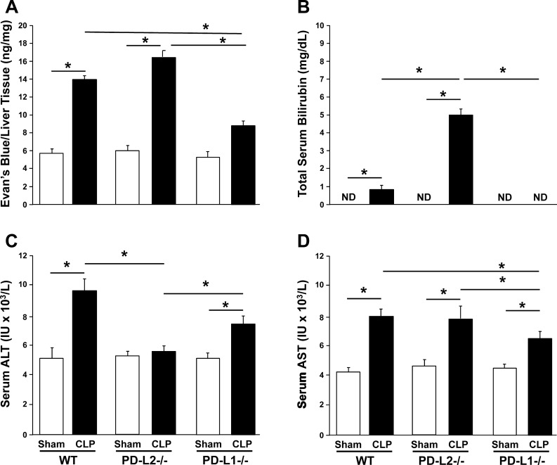
Fig. 2.
Indices of dysfunction and damage as a measure of septic mouse liver injury. Vascular permeability measured in nanograms of Evans blue dye over milligrams of liver tissue in wild-type (WT), programmed cell death receptor ligand 2 (PD-L2)−/−, and PD-L1−/− mice 24 h after cecal ligation and puncture (CLP) (n = 4/group) (A). Total blood bilirubin measured in mg/dL in WT, PD-L2−/−, and PD-L1−/− mice 24 h after CLP (n = 5/group) (B). Blood alanine aminotransferase (ALT) and aspartate aminotransferase (AST) measured in IU/L in WT, PD-L2−/−, and PD-L1−/− mice 24 h after CLP (n = 5/group) (C and D, respectively). Data are expressed as mean ± SE; *P < 0.05 by nonparametric Mann-Whitney U-test to evaluate P values when testing two subgroups. For multiple groups analysis, intergroup comparisons were performed by the Holm-Sidak test. ND, not detected.

