Figure 3.
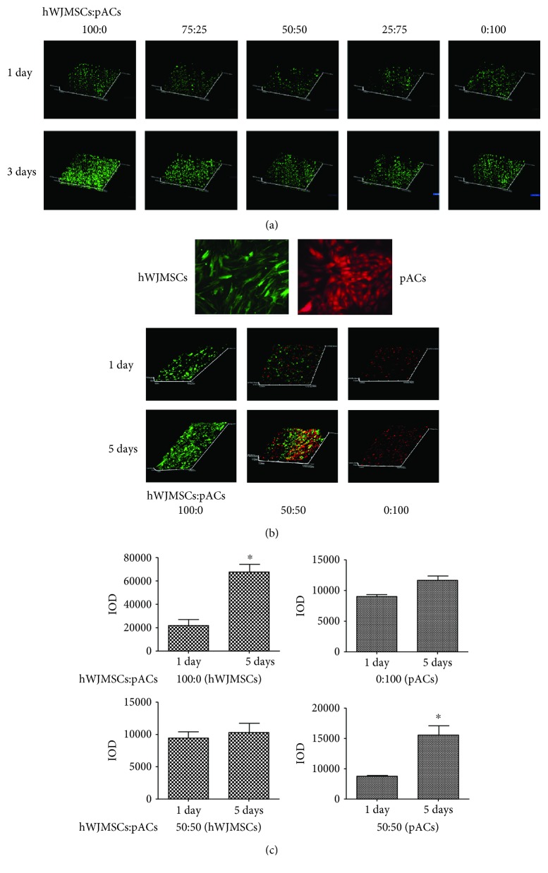
ACECM-oriented scaffold has good biocompatibility and the proliferation activity of pACs was significantly enhanced. (a) Dead/live staining of cell scaffold complexes using FDA/PI assay after coculturing for 1 day and 3 days by laser scanning confocal microscope, ×40; red color represents a dead cell and green color represents a living cell. (b) Green fluorescent lentivirus-labeled hWJMSCs, red fluorescent lentivirus-labeled pACs, laser scanning microscope, ×200; the proliferation assay was performed in three different groups (hWJMSCs : pACs), 100 : 0, 50 : 50, and 0 : 100, after coculturing for 1 day and 5 days; laser scanning confocal microscope, ×40. (c) 100 : 0 (hWJMSCs), IOD of hWJMSCs in the 100 : 0 group at 1 day and 5 days; 100 : 0 (pACs), IOD of pACs in the 100 : 0 group at 1 day and 5 days; 50 : 50 (hWJMSCs), IOD of hWJMSCs in the 50 : 50 group at 1 day and 5 days; 50 : 50 (pACs), IOD of pACs in the 50 : 50 group at 1 day and 5 days. The results are shown as means ± SD; n = 3, ∗ p < 0.05.

