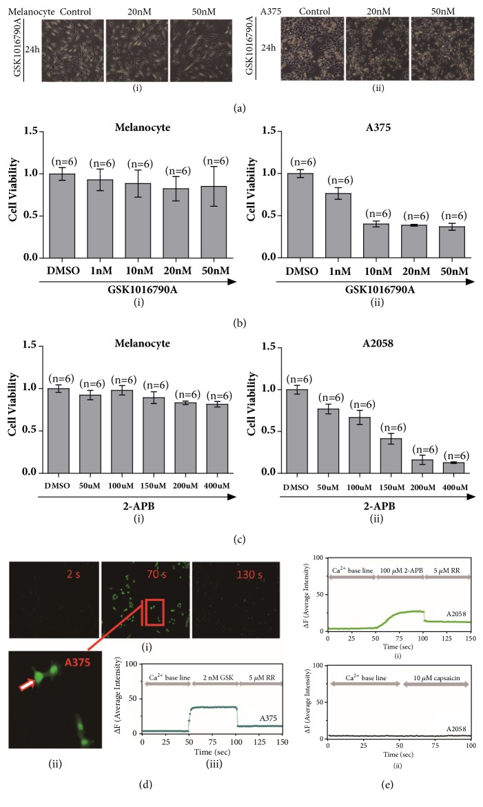
Figure 2.
Functional TRPVs mediated proliferation of melanoma cells. (a) Cells morphologically changed after GSK1016790A (20 nM and 50 nM) application to A375 melanoma cells and melanocytes via microscopy imaging (i) & (ii). (b) Cell viability test by MTT experiment exhibited inhibition of cell proliferation for melanoma A375 cells but not for primary epidermal melanocytes by treatment of either DMSO (control) or TRPV4 specific activator of GSK1016790A (1 nM, 10 nM, 20 nM, 50 nM) (i) & (ii). (c) Cell viability test by MTT experiment exhibited inhibition of cell proliferation for melanoma A2058 cells but not for primary epidermal melanocytes by treatment of either DMSO (control) or 2-APB (50 μM, 100 μM, 150 μM, 200 μM, 400 μM) (i) & (ii). (d) Measurement of calcium imaging in GSK1016790A application with 2 nM concentration in A375 melanoma cells was carried out, and intracellular calcium that dramatically enhanced was observed and calcium signal receded after a blocker of ruthenium red was applied (i). A representative single cell (ii) recording by time course showed calcium influx fluctuation before and after GSK1016790A application and channel blocker was added (iii), (n = 12, p < 0.01). (e) Measurement of calcium imaging showed 2-APB could induce calcium influx which could be blocked by TRP channel blocker of ruthenium red in A2058 cells (i), (n = 12, p < 0.01). When TRPV1 agonist of capsaicin was applied, there was no apparent calcium signal observed in A2058 melanoma cells (ii), (n = 19).

