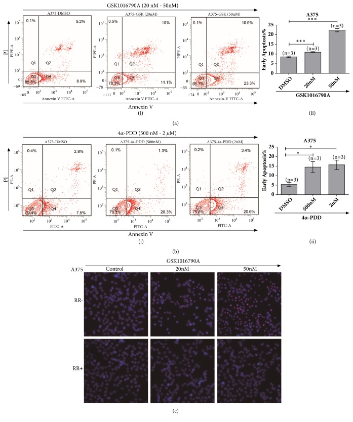
Figure 5.
GSK1016790A application induced apoptosis of human A375 melanoma cells. (a) Flow cytometry test via FITC-Annexin V and PI staining showed increasing apoptotic signals by treatment with GSK1016790A to A375 melanoma cells (i) & (ii). (b) Flow cytometry analysis via FITC-Annexin V and PI staining for 4α-PDD treatment exhibited enhancing apoptotic cells in melanoma A375 cells (i) & (ii). (c) Pretreatment of ruthenium red prominently attenuated apoptotic cells for the application of GSK1016790A with 20 nM and 50 nM to A375 melanoma cells; cells were stained with Hochest33342 and PI. All tests were performed in at least three independent experiments.

