
Figure 1.
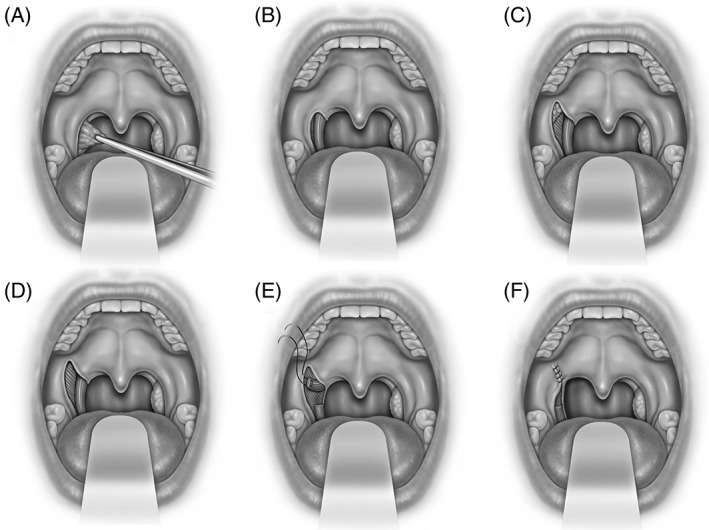
Expansion sphincterplasty is shown (A) tonsillectomy, (B) exposure of palatopharyngeus and superior constrictor muscles within the tonsillar fossa, (C) lateral palatal incision over lateral palatal space with exposure of supratonsillar fat, (D) removal of fat and fibers of palatoglossus provides exposure to superior constrictor and arching fibers of palatopharyngeus muscles, (E) palatopharyngeus muscle incised 1.5 cm. inferior to the fulcrum of rotation and the pedicle is sutured to pterygomandibular raphe or fibrous tissue lateral to the hamulus, and (F) mucosal closure.