
Figure 5.
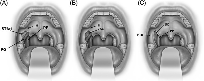
Anatomy of the lateral palatal space is shown. Exposure of palatoglossus muscle and supratonsillar fat (STfat) (in A), removal of supratonsillar fat in pterygoid humulus area (H) (in B) and palatoglossus muscle (PG) (in C) demonstrate the palatal anatomic relationships of the superior pharyngeal constrictor muscle (SC), palatopharyngeus muscle (PP), and pterygomandibular raphe (PTR).