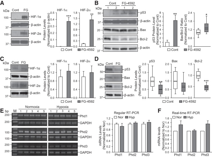
Fig. 7.
Increasing hypoxia-inducible factor (Hif) by pharmacologically blocking prolyl hydroxylase domain proteins (PHDs) upregulates p53 in human pulmonary arterial endothelial cells (PAECs) and mRNA expression level of PHDs is comparable in lung tissues from normoxic and chronically hypoxic mice. A: Western blot analysis of HIF-1α and HIF-2α in normal human PAECs treated with vehicle (Cont) and FG-4592 (FG, 100 µM for 48 h), a selective blocker of PHDs. Summarized data (means ± SE; n = 5; right) showing protein levels of HIF-1α and HIF-2α in control PAECs and FG-treated PAECs. B: Western blot analysis of p53, Bax and Bcl-2 in control PAECs and FG-treated PAECs. Summarized data (means ± SE, n = 5; right) showing the protein levels of p53 and the ratio of Bax/Bcl-2 in control PAECs and FG-treated PAECs. C: Western blot analysis of HIF-1α and HIF-2α in normal human PASMCs treated with vehicle (Cont) and FG-4592 (FG, 100 µM for 48 h). Summarized data (means ± SE, n = 5; right) showing protein levels of HIF-1α and HIF-2α in control pulmonary arterial smooth muscle cells (PASMCs) and FG-treated PASMCs. D: Western blot analysis of p53, Bax and Bcl-2 in control PASMCs and FG-treated PASMCs. Summarized data (means ± SE, n = 5; right) showing the protein levels of p53, Bax, and Bcl-2 in control PASMCs and FG-treated PASMCs. E: RNA was extracted and regular quantitative RT-PCR was performed to determine mRNA expression levels for Phd1, Phd2, and Phd3 (left). Summarized data (means ± SE, n = 5; right) showing the mRNA levels of Phd1, Phd2, and Phd3 in whole lung tissues from normoxia and hypoxia (10% O2 for 21 days) mice. F: summarized data (means ± SE, n = 5) represented mRNA levels for Phd1, Phd2, and Phd3 in whole lung tissues from normoxia and hypoxia (10% O2 for 21 days) mice, using real-time quantitative RT-PCR. For these studies, all cells used were between 5 and 8 passages. We compared the same passage number of cells for each experiment. *P < 0.05, **P < 0.01, and ***P < 0.001 vs. Cont or Nor.

