Fig. 7.
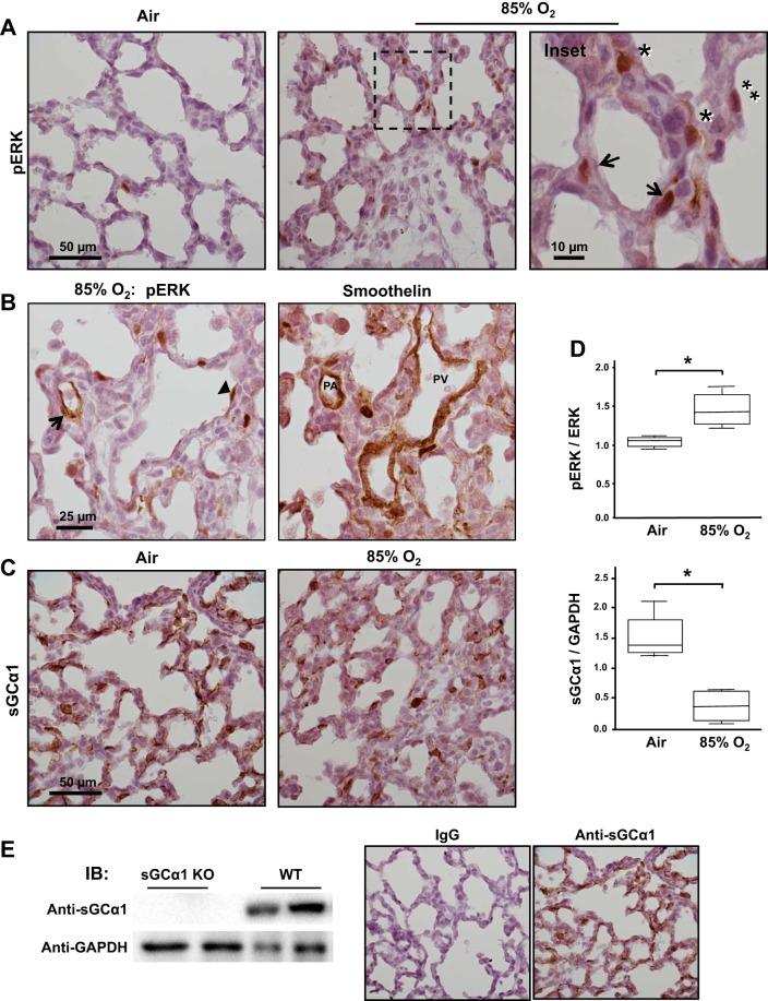
Lung injury increases ERK activation and decreases stimulated soluble guanylate cyclase (sGC) expression in the mouse pup. A: lung injury increases ERK phosphorylation in mouse pup parenchymal cells. Newborn mouse pups breathed either air or 85% O2 for 10 days, and then phosphorylated (p)-ERK protein was detected in lung tissue sections using immunohistochemistry and a colorimetric substrate (brown). The sections were counterstained with Gill’s hematoxylin. Cells in the blood vessel wall (arrows) and interstitial (*) and epithelial cells (**) are identified in the images, which are representative of lungs of 5 pups. B: p-ERK is detected in PASMC in the injured pup lung. p-ERK or smoothelin, a SMC marker protein, was detected in consecutive, 5-µm-thick sections of 85% O2-treated mouse pup lungs using antibodies and immunohistochemistry (brown). PASMC expressing p-ERK are identified (arrow) based on their localization within the blood vessel wall and reactivity with the anti-smoothelin antibody; an adluminal endothelial cell exhibiting ERK activation is also shown (arrowhead). The images are typical of 2 pups. C and D: pulmonary injury increases p-ERK and decreases sGCα1 protein expression in the newborn lung. sGCα1 protein expression was detected in air- or 85% O2-treated mouse pup lungs using an anti-sGCα1 antibody and immunohistochemistry (brown). sGCα1 and p-ERK protein expression were quantified in pup lung lysates using immunoblotting and densitometry; n = 4 each group; *P < 0.05. E: the anti-sGCα1 antibody specificity is shown by its inability to react with soluble proteins obtained from sGCα1-knockout (KO) in comparison with wild-type (WT) mouse pup lungs by immunoblotting. The secondary antibody specificity is supported by its lack of reactivity to WT pup lungs treated with rabbit IgG rather than the rabbit anti-sGCα1 antibody.

