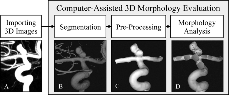
Figure 1:
Computer-assisted three-dimensional (3D) morphology workflow. The workflow includes: A) importing 3D images, to import 3D Digital Imaging and Communications in Medicine (DICOM) images of the intracranial aneurysm (IA); B) segmentation, to generate a 3D geometry of the intracranial aneurysm (IA); C) pre-processing, to isolate the region of interest and remove small vessel branches; D) morphologic analysis, to analyze the IA geometry and extract morphologic parameters.

