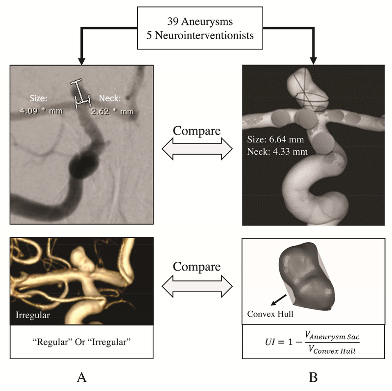
Figure 2:
Comparison of current clinical practice and computer-assisted 3D approaches for IA morphology evaluation. A) manual measurements of an internal carotid artery (ICA) aneurysm obtained from two-dimensional digital subtraction angiography (2D-DSA) and visual inspection of IA shape on 3D-DSA. B) Computer-assisted 3D size and neck measurements and quantification of shape of the same aneurysm using the undulation index (UI).

