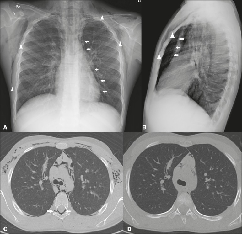
Figure 1.
Chest X-rays, in posteroanterior and lateral views (A and B, respectively), showing pneumomediastinum (arrows) and soft tissue emphysema (arrowhead). The lateral view better identifies the air delineating the mediastinum anteriorly (arrows). CT with an intermediate window, slices at the level of the bronchial bifurcation being acquired at admission (C) and 72 h later (D), showing free air delineating the mediastinal structures, bronchi, and pulmonary vessels, as well as pneumorrhachis (arrow in C). Note the significant improvement of the pneumomediastinum, subcutaneous emphysema, and pneumorrhachis at 72 h after the initial CT (D).

