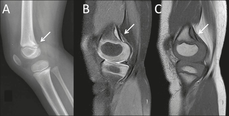
Figure 1.
A 4-year-old female with spontaneous knee pain. Note, as an incidental finding on an X-ray of the left knee (A), as well as in T2-weighted and T1-weighted MRI sequences of the same knee (B and C, respectively), a typical cortical desmoid (arrows) in the distal femoral metaphysis, located in the posterior cortex.

