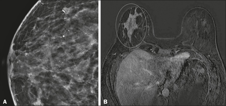
Figure 4.
A: Magnified craniocaudal mammography showing extensive grouping of grossly heterogeneous microcalcifications with segmental distribution in the right breast, together with a metallic clip (tissue marker) placed during the biopsy. B: Axial MRI sequence of the breasts, with digital subtraction, showing an area of heterogeneous non-nodular enhancement with segmental distribution and minimal post-contrast enhancement, occupying the lower outer quadrant of the right breast, corresponding to the findings described in the mammographic examination. The patient underwent mastectomy, and the pathology study revealed DCIS, nuclear grade 3, invading the lobules.

