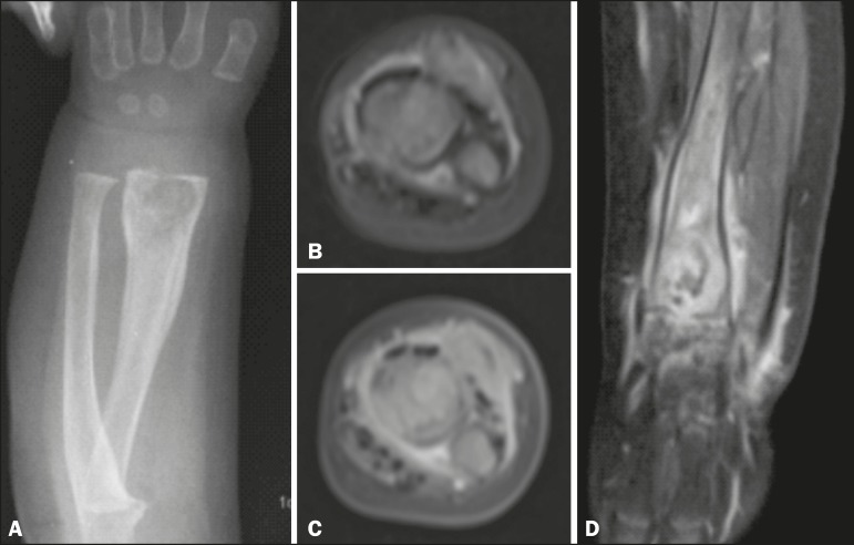
Figure 1.
A: Anteroposterior X-ray of the forearm. Round osteolytic formation with partially defined margins, cortical irregularity, and periosteal reaction in the distal third of the radius. B: Axial proton density-weighted MRI. Expansile ill-defined solid heterogeneous lesion in the bone marrow of the distal metaphysis of the radius. Note the linear image with a hyperintense signal in the metadiaphysis and cortical discontinuity suggestive of fracture. C: Contrast-enhanced axial T1-weighted MRI with fat suppression. The signal intensity is similar to that of cartilaginous tissue, with hyperintense foci. D: Contrast-enhanced coronal T1- weighted MRI with fat suppression. Note that the lesion focally extends beyond the physis and infiltrates the perilesional soft tissue, with significant gadolinium enhancement, persistence of small loculated lesions with hypointense signals, and fluid infiltration, as well as enhancement of the joint spaces, muscle, and subcutaneous tissue.

