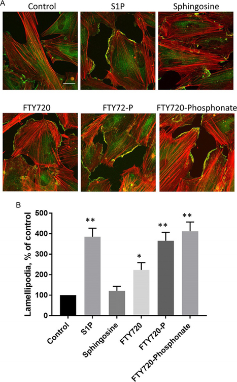
Fig. 3.
Effects of sphingolipids on endothelial lamellipodia formation. (A) Human lung microvascular endothelial (HLMVECs) cells were treated with vehicle, S1P (1μM), sphingosine (1μM), FTY720 (1μM), FTY720-P (1μM), or FTY720-phosphonate (1μM), for 30min. Cells were subjected to immunofluorescent staining for cortactin (green) and actin (red). Images were obtained with Zeiss LSM 880 confocal microscope, 63 × oil objective, scale bar=20μm. (B) Statistical analysis of lamellipodia formation presented as percentage of control, and at least 20 cells were analyzed for each condition. *P<0.05 vs control; **P<0.01 vs control.

