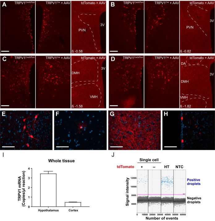
Fig. 1.
Expression of transient receptor potential vanilloid type 1 (TRPV1) in the dorsomedial hypothalamic nucleus (DMH) and the paraventricular nucleus of the hypothalamus (PVN). A–D: representative images demonstrate TRPV1-expressing neurons in PVN (A and B) and DMH (C and D) in TRPV1Cre/tdTom mice (left), in TRPV1Cre mice injected with a Cre-dependent adeno-associated virus (AAV) construct (center), and in control tdTomato mice injected with the same viral construct (right). E–H: representative images show TRPV1 expression in neurons (E) and astrocyte-like cells (F) in the DMH, dense processes in the ventromedial hypothalamus (G), and ependymal-like cells lining the third ventricle (H). I: TRPV1 mRNA in the hypothalamus and cortex in adult mice. J: expression of TRPV1 in single cells. Droplet digital PCR was used to reveal TRPV1 expression in single cells. Positive droplets containing a single copy of TRPV1 cDNA (blue dots) were identified in tdTomato-expressing neurons and in diluted hypothalamic mRNA (HT; positive control), which demonstrates TRPV1 expression in tdTomato fluorescent neurons. There was no positive droplet in tdTomato-negative neurons or in the no-template control (NTC; negative control). Negative droplets (black dots) are droplets that do not contain TRPV1 cDNA. DA, dorsal hypothalamic area; VMH, ventromedial hypothalamic nucleus; 3V, 3rd ventricle. β, distance from bregma; tdTomato +, tdTomato-positive neuron; tdTomato, nonfluorescent neuron. Scale bars: 100 µm (A–D), 50 µm (E–H).

