Fig. 3.
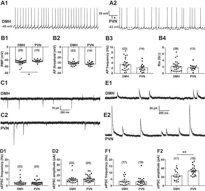
Basic cellular properties of transient receptor potential vanilloid type 1 (TRPV1)-expressing neurons in the dorsomedial hypothalamic nucleus (DMH) and the paraventricular nucleus of the hypothalamus (PVN). A: representative recordings from TRPV1-expressing neurons in the DMH (A1) and the PVN (A2). B: cellular properties of TRPV1-expressing neurons in the DMH and the PVN. B1: resting membrane potential (RMP). B2: threshold of action potentials (APs). B3: frequency of APs. B4: input resistance (Rin). C: representative recordings of spontaneous excitatory postsynaptic currents (sEPSCs) at holding potential of −60 mV in the DMH (C1) and the PVN (C2). D: frequency (D1) and amplitude (D2) of sEPSCs in TRPV1Cre/tdTom neurons in the DMH and the PVN. E: representative recordings of spontaneous inhibitory postsynaptic currents (sIPSCs) at holding potential of 0 mV in the DMH (E1) and the PVN (E2). F: frequency (F1) and amplitude (F2) of sIPSCs in TRPV1Cre/tdTom neurons in the DMH and the PVN. Numbers of recorded cells are shown in parentheses. *Significance (P < 0.05); **significance (P < 0.01).

