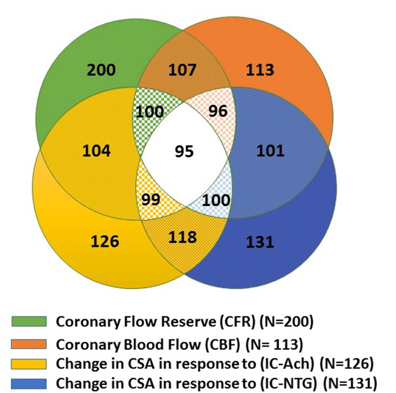
Figure 1. Distribution of selected coronary reactivity testing in 224 women with signs and symptoms of ischemia.
Most women underwent evaluation of more than one coronary reactivity pathway. Green color represents women who underwent evaluation of non-endothelium dependent microvascular reactivity using coronary flow reserve (CFR); orange color represents women who underwent evaluation of endothelium dependent microvascular reactivity using coronary blood flow (CBF); yellow color represents women who underwent evaluation of endothelium dependent epicardial coronary reactivity using change in coronary artery cross sectional area in response to intracoronary acetylcholine (IC-Ach); while blue color represents women who underwent evaluation of non-endothelium dependent epicardial coronary reactivity using change in coronary artery cross sectional area in response to intracoronary nitroglycerine (IC-NTG).

