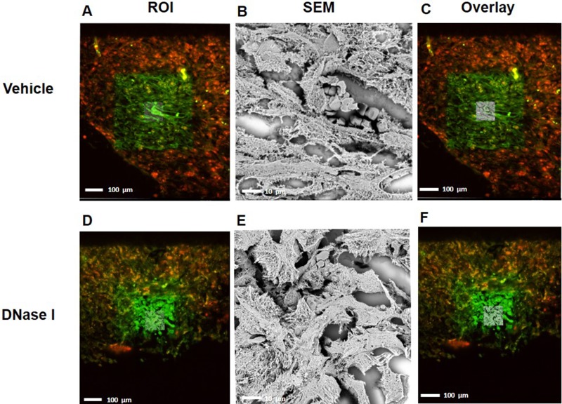
Figure 3. CLEM images indicating that NETs co-localized with murine colon cancer metastasis tissue.
(A, D) selected region of mouse GFP labeled-tumor tissue (green) containing citrullinated histone 3 (H3Cit-red) from vehicle-treated group and DNase1 treated group (B, E) Scanning electron microscope of tumor tissue shows web-like NET structure and (C, F) overlay of region of interest with SEM. ROI; Region of Interest, SEM; Scanning Electron Microscope.

