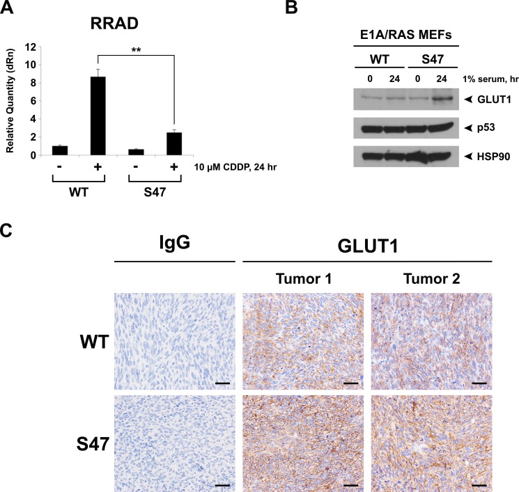
Figure 2. Decaresed induction of RRAD and increased expression of GLUT1 in cells and tumors containing the S47 variant.
(A) WT and S47 E1A/RAS MEFs were treated with 10 μM Cisplatin (CDDP) for 24 hours. Cellular RNA was isolated and used for quantitative reverse transcription-PCR analysis, and levels of RRAD were normalized to cyclophilin A. Values shown reflect the mean ± standard deviation of technical replicates. Shown are representative data of two independent clones of each genotype. (B) Western blot analysis of GLUT1, p53 and HSP90 (control) in WT and S47 E1A/RAS MEFs subjected to nutrient-deprived conditions (1% serum) for 24 hours. (C) GLUT1 protein staining in formalin fixed paraffin embedded tumors from WT and S47 E1A/RAS cells. Staining using Rabbit IgG was used as a negative control (scale bar, 100 µm). Shown are representative data from 5 independent tumors of each genotype.

