Figure 2.

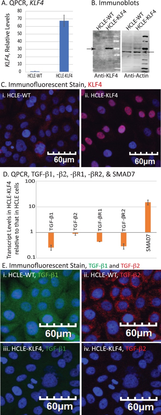
Overexpression of KLF4 in HCLE cells alters the expression levels of TGF-β signaling pathway components. (A) qPCR showing increased levels of KLF4 mRNA in HCLE-KLF4 compared with HCLE-WT. (B) Immunoblot showing overexpressed KLF4 in HCLE-KLF4 cells. (C) Immunofluorescent stain showing increased expression and nuclear localization of KLF4 in HCLE-KLF4. (D) HCLE-KLF4 cells show decreased levels of TGF-β1, -β2, -βR1, and -βR2 and increased expression of TGF-β signaling inhibitor SMAD7. Data are presented in logarithmic scale to accommodate large range in values. Data show results from three independent experiments performed in triplicates and reported as means ± SEM. (E) Immunofluorescent stain confirming the increased expression of TGF-β1and TGF-β2 in HCLE-KLF4 (iii, iv) compared with HCLE-WT cells (i, ii). Images were acquired at 20×; scale bar: 60 μm.
