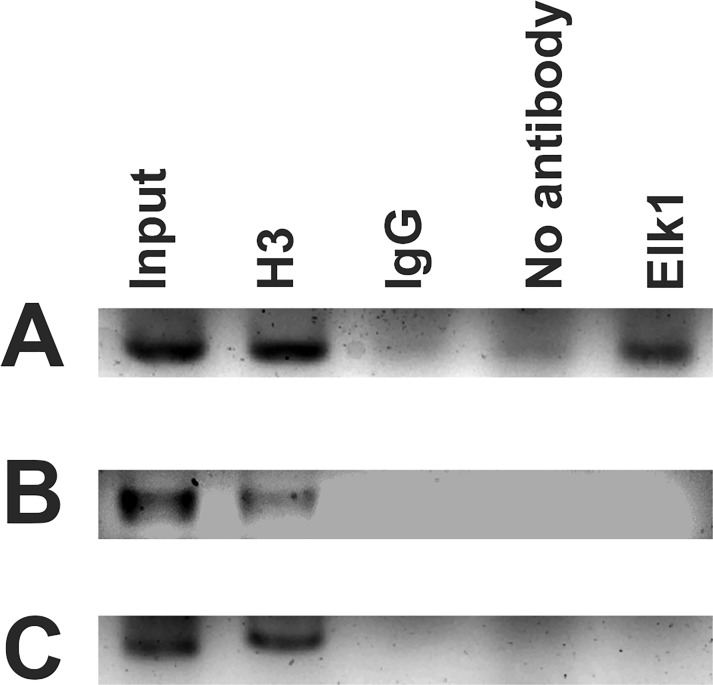
Fig 3. PCR results of ChIP assay.
ChIP assay was performed with SH-SY5Y cells. 2% input DNA; H3 (positive control), rabbit IgG (negative control), no antibody (beads only, negative control), and Elk1 precipitated reactions were amplified with specific primers for each binding sites separately. ChIP results for +113/+122 binding site (A), +253/+262 binding site (B), and -428/-419 binding site (C) are indicated.

