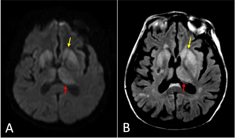
Figure 2. Axial magnetic resonance imaging (MRI) of Case 2.
(A) Diffusion-weighted image (DWI) at the level of the basal ganglia demonstrating asymmetrically increased signal primarily in the striatum (yellow arrow) and medial and posterior thalamus (red arrow) reminiscent of the “hockey stick” sign frequently seen in variant Creutzfeldt-Jakob disease (CJD). (B) T2-weighted fluid attenuation inversion recovery (T2-FLAIR) image with less corresponding hyperintense signal.

