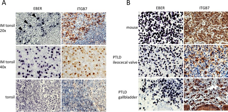
FIG 2.
Expression of integrins and localization of EBV-infected B cells. (A) Consecutive histological sections of a tonsil removed from a patient with IM was stained for EBER (top left) or ITGB7 (top right) (n = 7). The top and middle pictures were taken at low (×20) and high (×40) power. (Bottom) Similar investigations performed in the tonsil of a patient without EBV acute infection (n = 4). (B) The expression of ITGB7 in various EBV-infected tumors from humans or immunosuppressed mice was assessed by immunohistochemistry (n = 4 and n = 8, respectively).

