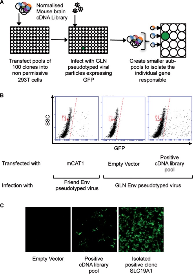
FIG 1.

cDNA library screen to identify the GLN cellular receptor. (A) Schematic representation of the screening method. Nonpermissive 293T cells were transfected with pools of 100 cDNAs from a normalized mouse brain library and then exposed to GFP-expressing GLN-pseudotyped virions 48 h later. Seventy-two hours after viral exposure, the 293T cells were analyzed for GFP expression by fluorescence microscopy and flow cytometry. Positive pools were retransformed into competent bacteria and used to generate subpools until the unique positive clone was identified. (B) Flow cytometric analysis of the cDNA screen showing the cDNA pool containing the GLN receptor. Cells transfected with mouse CAT-1 (mCAT1) and infected with Friend Env-pseudotyped viruses were used as positive controls for the assay. SSC, side scatter. (C) Fluorescence microscopy images of GFP-positive 293T cells after infection with GLN-pseudotyped viruses when transfected with the positive cDNA pool and the isolated cDNA (SLC19A1) responsible for enabling infection.
