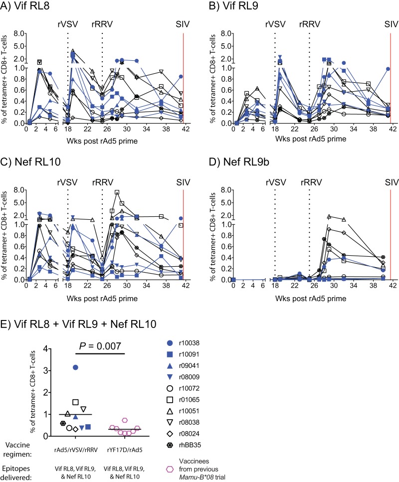
FIG 2.
Kinetics of vaccine-induced CD8+ T-cell responses targeting Mamu-B*08-restricted SIV epitopes. Fluorochrome-labeled Mamu-B*08 tetramers folded with peptides corresponding to SIV epitopes were used to track vaccine-elicited SIV-specific CD8+ T cells in PBMC. (A to D) The percentages of live tetramer+ CD8+ T cells specific for Vif RL8 (aa 172 to 179) (A), Vif RL9 (aa 123 to 131) (B), Nef RL10 (aa 137 to 146) (C), and Nef RL9b (aa 246 to 254) (D) are shown at multiple time points throughout the vaccine phase. The times of each vaccination (black dotted lines) and the day of the first IR SIVmac239 challenge (red solid line) are indicated in each graph. (E) Comparison of the total magnitude of vaccine-induced CD8+ T cells against Vif RL8, Vif RL9, and Nef RL10 at the time of the first SIV challenge between vaccinees in the present experiment and those in our previous Mamu-B*08 SIV vaccine trial (36). The P value was determined by Student's t test after log transformation. Lines represent means, and each symbol denotes one vaccinee. The vaccinees in the present experiment that resisted greater than 6 IR SIV challenges (r10038, r10091, r09041, and r08009) are color coded in blue. The remaining six vaccinees that acquired SIV infection and manifested partial or no control of viral replication are indicated by black symbols.

