FIG 9.
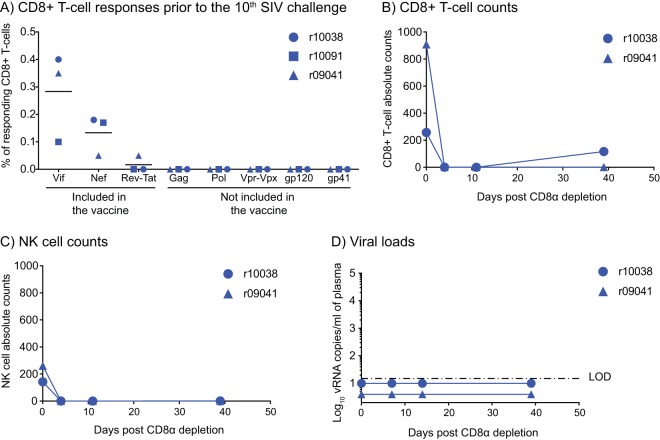
No evidence for de novo induction of SIV-specific CD8+ T-cell responses or SIV rebound following CD8α depletion in vaccinees that resisted repeated IR exposures to SIV. (A) Macaques r10038, r10091, and r09041 were bled 4 days prior to the 10th IR SIV challenge, and an ICS assay was carried out in PBMC. The antigen stimuli for this assay consisted of peptides corresponding to SIV antigens that were included in the vaccine (Vif, Nef, and Rev-Tat) as well as proteins that were not included in the vaccine (Gag, Pol, Vpr, Vpx, and Env). The percentages of responding CD8+ T cells displayed in the panel were calculated by adding the background-subtracted frequencies of positive responses producing any combination of IFN-γ, TNF-α, and CD107a. Lines represent means, and each symbol corresponds to one vaccinee. (B to D) To evaluate whether r10038 and r09041 harbored replication-competent SIV, these animals were treated with a single i.v. infusion of 50 mg/kg of a CD8α-depleting MAb 39 weeks after the 10th IR SIVmac239 challenge. Monkey r10091 was not subjected to this procedure because it had to be euthanized prior to it. The absolute counts of CD8+ T cells (CD3+ CD8α+) and NK cells (CD3− CD8α+ CD16+) per microliter of blood after the CD8α depletion are shown in panels B and C, respectively. (D) Viral loads after CD8α depletion. The VLs were log transformed and correspond to the number of vRNA copies per milliliter of plasma. The dash-dot line indicates the limit of detection (LOD) of the VL assay (15 vRNA copies/ml of plasma).

