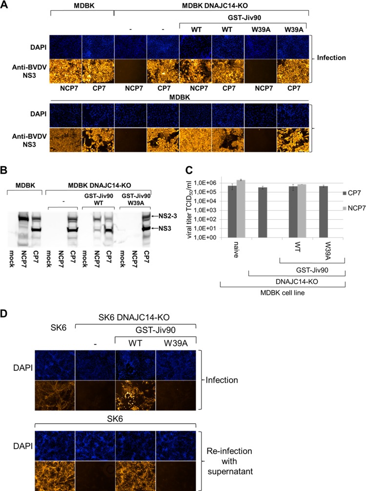
FIG 6.
Expression of GST-Jiv90(WT) functionally rescues the DNAJC14 gene knockout in MDBK and SK6 cells and supports noncp pestivirus replication in MDBK and SK6 DNAJC14-KO rescue cells. (A, upper) Immunofluorescence analysis after infection of different target cells. Naïve MDBK, MDBK DNAJC14-KO, MDBK DNAJC14-KO GST-Jiv90(WT), and MDBK DNAJC14-KO GST-Jiv90(W39A) rescue cells were infected with CP7 and NCP7 viruses at an MOI of 0.1. At 72 hpi, immunofluorescence analysis was performed using a monoclonal anti‐BVDV NS3 antibody. Nuclei are DAPI stained. (A, lower) Immunofluorescence analysis of the reinfection experiment. Cell culture supernatants from CP7 or NCP7 infection of naive MDBK and MDBK DNAJC14-KO cells as well as MDBK DNAJC14-KO GST-Jiv90(WT) and MDBK DNAJC14-KO GST-Jiv90(W39A) rescue cells were used to infect naive MDBK cells with 500 μl of filtered supernatant. At 72 hpi, immunofluorescence analysis was performed using a monoclonal anti‐BVDV NS3 antibody. Cell nuclei were counterstained with DAPI. (B) Western blot analysis of NS2-3 cleavage in MDBK and MDBK DNAJC14-KO cells as well as MDBK DNAJC14-KO GST-Jiv90(WT) and MDBK DNAJC14-KO GST-Jiv90(W39A) rescue cells infected with noncp BVDV NCP7 and cp BVDV CP7. The lysate separated in each lane represents about 5 × 10⁵ cells. Lysates were prepared at 72 hpi. Positions of NS3 and NS2-3 are marked with arrows. Depicted is a representative Western blot of three independent experiments. (C) Viral titer analyses of MDBK and MDBK DNAJC14-KO cells as well as MDBK DNAJC14-KO GST-Jiv90(WT) and MDBK DNAJC14-KO GST-Jiv90(W39A) rescue cells infected with noncp BVDV NCP7 and cp BVDV CP7. Cell culture supernatants collected at 72 hpi (MOI of 0.1) were analyzed for infectious virus by limiting dilution assay (TCID50/ml). Mean values and standard deviations of viral titers from three experiments are depicted. (D, upper) Naïve SK6 and SK6 DNAJC14-KO cell as well as SK6 DNAJC14-KO GST-Jiv90(WT) and SK6 DNAJC14-KO GST-Jiv90(W39A) rescue cells were infected with CSFV Alfort-Tübingen at an MOI of 0.1. At 72 hpi, immunofluorescence analysis was performed using a monoclonal anti‐CSFV NS3 antibody. Nuclei are DAPI stained. (D, lower) Immunofluorescence analyses of reinfection experiment with naive SK6 cells. Cell culture supernatants from CSFV infection of naive SK6 and SK6 DNAJC14-KO cells as well as SK6 DNAJC14-KO GST-Jiv90(WT) and SK6 DNAJC14-KO GST-Jiv90(W39A) rescue cells were used to infect naive SK6 cells with 500 μl of filtered supernatant. At 72 hpi, immunofluorescence analysis was performed using a monoclonal anti‐E2 antibody. Cell nuclei were counterstained with DAPI. Depicted is a representative data set from three independent experiments.

