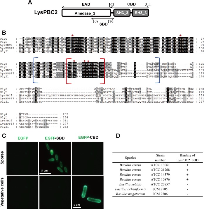
FIG 1.
Spore binding domain of LysPBC2. (A) Schematic diagram of the multimodular structure of LysPBC2. (B) Sequence alignment of LysPBC2-related endolysins. Conserved residues are shaded in gray (>60% identity) and black (>80% identity). *, conserved amidase catalytic residue. The brackets indicate SBD (blue) and SBD core (red) regions. (C) Binding capacities of EGFP only, EGFP-SBD, and EGFP-CBD for B. cereus spores and vegetative cells. (D) Spore binding selectivity of the SBD.

