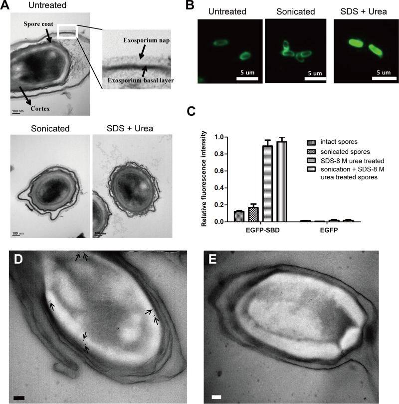
FIG 2.
The outermost layer of the cortex is the most probable binding target of the SBD. (A, top) TEM of untreated spores. Inset shows exosporium basal layer and hair-like nap. (Bottom) TEM of spores sonicated or treated with SDS and urea buffer. All panels stained with ruthenium red to visualize the heavily glycosylated exosporium nap. (B) Binding profiles of EGFP-SBD with B. cereus intact spores, sonicated spores, and SDS–urea-treated spores. (C) Relative fluorescence intensities of differently treated spores after incubation with EGFP-SBD and EGFP-only proteins. Immunogold electron micrographs of ultrathin sections of B. cereus spores were labeled with primary antibody (D) or without primary antibody (E). The arrows point to 10-nm gold nanoparticles. The scale bars represent 100 nm.

