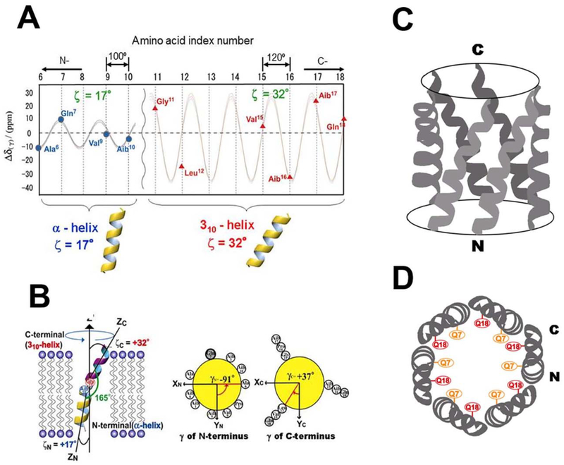
Fig. 8.
(A): Chemical shift oscillation patterns of alamethicin. Chemical shift oscillation curves were obtained from the chemical shift anisotropies of the N-terminus (AIa6, Gln7, Val9 and Aib10) and the C-terminus (Val15, Aib16, Aib17, and Gln18). The tilt angles of the N- and C-termini were determined to be 17° and 32°, respectively. The dihedral angles of the peptide planes between the n− and n + 1-residues of the N- and C-termini were determined to be α- and 310-helices, respectively. (B): Structure and topology of alamethicin bound to a DMPC bilayer, as determined from chemical shift oscillation data [50]. Side view (C) and top view (D) of the hexameric oligomer of alamethicin in the membrane environment.

