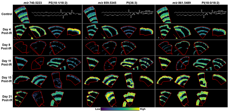
Figure 3.

MALDI-MS images of PE’s and PI’s localized to the crypt-villi axis in control and at days 4, 8, 11, 15 and 21 post-exposure. Example lipid structures are given, double bond positions are unknown, and PI(36:3) is a tentative structure based on the more commonly reported acyl chains for this species.
