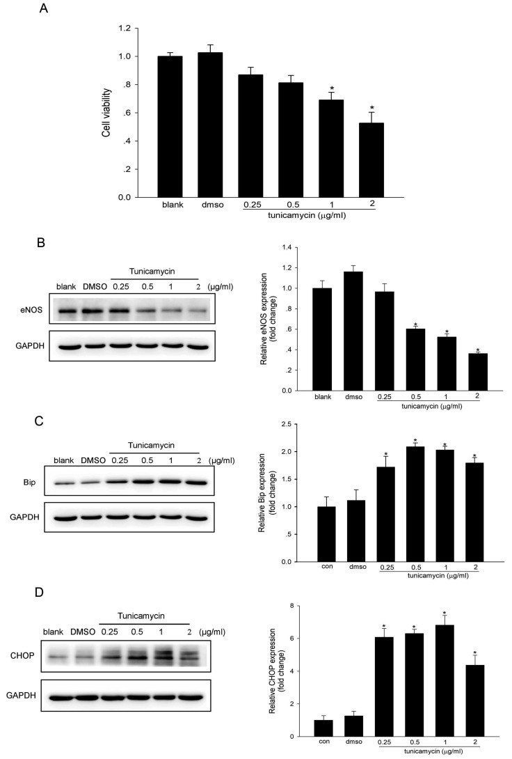
Fig. 1. Tunicamycin induced endothelial dysfunction by ER stress.
HUVECs were treated with 0.25, 0.5, 1, 2 µg/ml tunicamycin for 24 h. Cell viability was assayed with Cell Counting Assay Kit-8 (A). Expression of eNOS (B), BiP (C) and CHOP (D) was determined by western blotting. GAPDH was used as an internal control. *p < 0.05 vs. control; n = 4.

