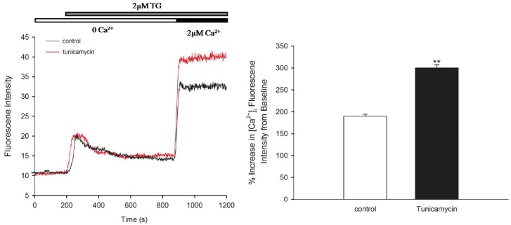
Fig. 2. Alteration of intracellular Ca2+ concentration in HUVECs induced by tunicamycin.
HUVECs were treated with 1 µg/ml tunicamycin for 24 h. Cells were incubated in a Ca2+-free buffer containing 2 µM thapsigargin for 8 min, and Ca2+ influx was induced during the Ca2+ loading period (left panel). Ca2+ influx was enhanced by tunicamycin (right panel). **p < 0.01 vs. control; n = 60 cells.

