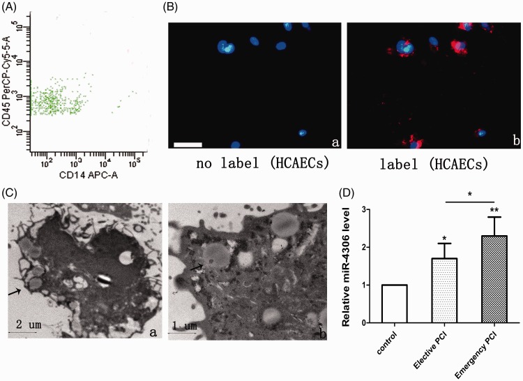
Figure 1.
EVs from patients’ HMDMs were taken up by HCAECs. (A) Representative flow cytometry analyses of the EV population derived from HMDMs. (B) Fluorescently-labelled EVs entering HCAECs. Fluorescently-labelled HM-EVs were incubated with HCAECs for 0 (a) or 12 hours (b) at 37°C. Original magnification, ×100; scale bar, 50 µm. (C) Electron microscopy shows that HCAECs (a, b) engulf HM-EVs (arrows). (D) Levels of miR-4306 in HM-EVs in control patients, elective patients, and emergency patients (n = 4). *p < 0.05, **p < 0.01. EV: extracellular vesicle; HM: human monocyte; HMDMs: human monocyte-derived macrophages; HCAECs: human coronary artery vascular endothelial cells; miRNA: microRNA.

