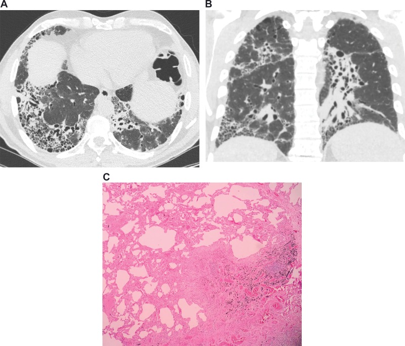
Figure 2.
Images from a 53-year-old male with idiopathic pulmonary fibrosis. (a) Axial and (b) coronal computed tomography images demonstrating areas of honeycombing, reticulation and subpleural predominance. (c) Histopathology images demonstrating areas of marked fibrosis, with architectural distortion and fibroblast foci, alternating with areas of normal parenchyma.

