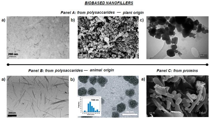
Figure 1.
Morphological characterization of bio-based nanofillers. Panel A: Polysaccharides—plant origin: (a) Transmission Electron Microscopy (TEM) image of Cellulose Nanocrystals CNC [27]; (b) Field Emission Scanning Electron Microscopy (FESEM) image of lignin nanoparticles [34]; (c) TEM image of starch nanoparticles [35]. Panel B: Nanofillers from Polysaccharides—animal origin: (a) TEM image of Chitin nanocrystals [36], (b) TEM image of modified chitosan nanoparticles (CSNP) by poly (ethylene glycol) methyl ether methacrylate (PEGMA) (PEGMA-graft-CSNP) [37]. Panel C: From proteins: (a) Scanning Electron Microscopy (SEM) image of nanokeratin [38].

