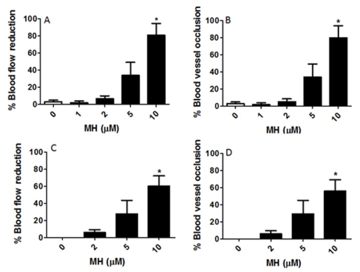
Figure 3.
Reduction of blood flow and vascular occlusion by MH. Embryos were exposed to 1, 2, 5, and 10 μM MH from 5 h post fertilization (hpf) to 6 dpf (0–6 dpf, (A,B)) or from 2 dpf to 6 dpf (2–6 dpf, (C,D)). This figure showed that the reduction in blood flow was proportionally correlated with blood vessel occlusion. (A) Blood flow was observed on days 2, 3, 4, 5, and 6 to verify the duration of the reduction of blood flow. This panel exemplifies the percent reduction in blood flow until 10 dpf in response to various concentrations of MH for 0-6 dpf. (B) This panel shows percent occlusion following MH treatment. During the treatment, blood flow was significantly reduced compared to the control immediately after occlusion. (C) Thirty-two embryos (2 dpf) were randomly divided into four groups (n = 8). Embryos were treated with the indicated concentrations of MH and 0.02% DMSO (control) in embryo medium and were maintained at 26 ± 1 °C for 4 days. This figure shows reduced blood flow in MH-treated embryos (2–6 dpf). (D) This figure demonstrates a blockage that prevents normal flow of blood for the MH-treated embryos exposed to various concentrations of MH from 2 dpf to 6 dpf. Each bar represents data pooled from 4–5 independent experiments. Statistical analysis was performed by one-way analysis of variance (ANOVA) followed by Tukey’s post-hoc multiple comparison test. p < 0.05 was considered as significant. The asterisk (*) indicates values significantly different from the control.

